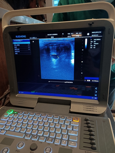
Veterinary ultrasound waves are transmitted through high-frequency sound waves. Its frequency is 20-20000 Hz. When waves collide with tissues, liquids, or gases, some waves are absorbed and then captured by ultrasound equipment and transmitted through images.
The depth of the echo determines the maximum depth at which the organization is displayed on the monitor. The results are expressed in decibels (dB), indicating the signal intensity pointing to the tissue to be ultrasound examined. Adjustments must be made according to the thickness of the fabric. Veterinarians recommend using lower power to achieve good results in images.
The most popular ultrasound in the market currently is electronic models for real-time analysis, which can image the content being analyzed in real time.
In order to generate the best image, it is necessary to find sensors with a frequency of 5 MHz, as they can effectively lock in depths up to 15 centimeters for spleen, kidney, liver, gastrointestinal and reproductive analysis.
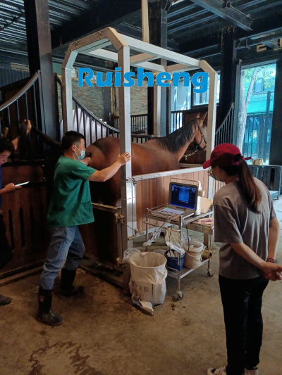
One of the most commonly used analyses currently is ultrasound, which is applied in the diagnosis of soft tissue diseases in the limbs of horses. That's why conducting analysis requires extensive knowledge from veterinarians.
Post time: Apr-20-2023